Research
High Resolution 3D Functional Anatomy Database of the Kidney
The kidney’s vascular structure is shaped by its role in blood filtration. In particular, its form interacts with the tubular structure through features such as the renal corpuscle for filtration or countercurrent multiplication loops for water and ion reabsorption.
This project aims to elucidate the full three-dimensional structure of the renal vasculature in its entirety down to the smallest capillaries, together with the entire urinifereous system, as well as the location of the renal EPO-producing cells. To this end, sample preparation methods will be developed to allow the necessary structural imaging data to be gathered using X-ray micro-computed tomography (µCT) and 3D light microscopy.
The obtained structural data are expected to give new insights into the interplay of the various structural features of the kidney on an organ-wide level and enable us to improve computational models simulating kidney functions and properties.
The imaging protocols developed are not limited to the kidney and should allow for the acquisition of similar structural data for a wide variety of tissues, both for regular anatomy and disease models.
Downloads
The following video showcases an X-ray micro-CT dataset of a right mouse kidney perfused with a radiopaque plastic resin mixture.
Highlights
To develop a protocol that allows for high resolution multi-modal imaging of organs and large tissue samples.
To construct a high resolution 3D functional anatomy database of the mouse kidney.
-
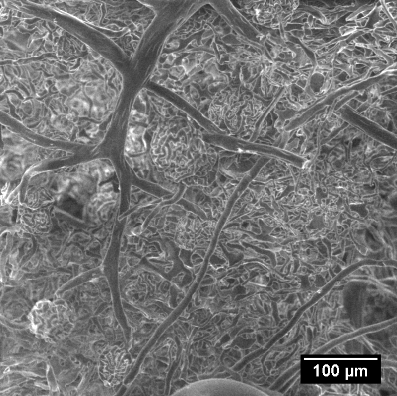
Figure 1
-
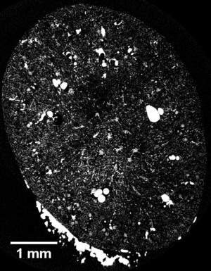
Figure 2